Free alternatives to Pathomation

Evil money-grabbing thieves

Pathomation is a for-profit company. We pride ourselves in offering various commercial products. Over the years, we’ve developed a comprehensive software platform for digital pathology and virtual microscopy. This platform consists of various components, which can be purchased, rented, or even licensed.

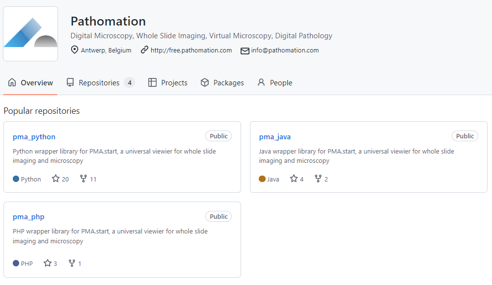
We strive to contribute to the Open Source community where we can. Some of our developers get referenced occasionally in papers, and our SDKs are publicly available through GitHub. On occasion, we contribute to Open Source projects ourselves.

Sample slides

Occasionally, people also ask us about where to find sample slides.

A great inventory of publicly available whole slide images can be found at both OpenSlide and the OMERO project:

What else do you need?

Oh yes, a way to actually read these data!

Reading WSI data

There are two well established Open Source projects for WSI data and virtual microscopy:

As you’re getting started adding digital pathology features to your own software, there are other routes to explore, too:

  • Work with slide scanner vendors
  • Adapt or translate code from BioFormats
  • Adapt or translate code from OpenSlide

All of these have downsides however:

  • Scanner vendors: You can start negotiating with any particular scanner vendor to adapt their proprietary SDK. Congrats; after months of lobbying work, you’ll have the (rather unpleasant) job of tasking one of your engineers to incorporate a new arcane DLL into your code, for a single framework (so expect to repeat this process over and over again for the next couple of years as your platform gains broader adoption)
  • Bioformats: is open source, so you could learn from their code and translate parts of it into your own. You can try it at least. Bioformats is a Java stack. If you can integrate that directly, you’re lucky. You could possibly transcribe the original Java-code to your own platform of course (C#, C++, Python, Rust, Haskell, Martian…). Same thing here: you’ll have the (still rather unpleasant, IMHO) task of tasking one of your engineers to actually do all of this, for a single file format (and again expect to repeat this process over and over again for the next couple of years as your platform gains broader adoption)
  • OpenSlide: lots of support, lots of bindings, but a bit outdated (limited file format support), limited functionality (not a server, no concept of “tiles”, …), and not adapted for today’s regulatory environment (GAMP, CE-IVD(R), 21CFR.11, …).

Of course all of this might change in the future. But as you sit and wait, we are here to help now, and  you may already consider what Pathomation has to offer as an alternative:
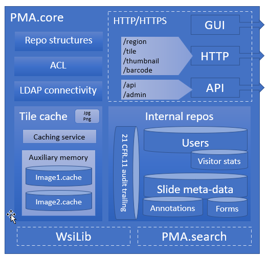
PMA.start

A central theme in our platform is the PMA.core tile server. A light-weight version of PMA.core is available free of change as PMA.start through https://free.pathomation.com.

PMA.start (and the entire Pathomation software stack by extension) supports any number of native file formats. So, you have to choices to go with here actually:

The advantage of this approach? Once you’re in the DICOM or TIFF world, you’re pretty much independent from any proprietary software vendor. Use Paint.Net to make snapshots of particular Regions of Interest (ROIs), use Photoshop to run a sequence of filters to detect hormone receptors in breast biopsies.

The downside: this doesn’t scale particularly well with large datasets or high throughput.

It is our view then that reading native file formats directly will most likely remain the most efficient way to offer digital pathology services in the foreseeable future.

The hidden cost of free software

Our point with all the above is: free is not free. You can always download free software, but after 10 years I daresay it’s NEVER going to do what you actually need it to do. But you’ll need to invest R&D, write your own unit and integration tests, add additional regulatory and compliance layers, combine development stacks and frameworks (the proverbial round peg in square hole)… Assuming that you have these people in-house, or can find them: these DON’T work for free.

Last but not least, there’s an opportunity cost here as well: again, if you have super-dev-guy/girl; don’t you want him/her to work on the core mission of your company (like saving patients’ lives, or developing awesome eXplainable AI)?

Plan ahead if you can

We’re not against Open Source software. We don’t advocate a radical choice between the two. We think this can be an inclusive and/and story, where both models can live in harmony.

We do think however that in many scenarios, Pathomation is the right (and even the better) course of action from the start. Adapt PMA.start early on in your projects as an individual single user solution. Then, as you learn more and expand the scope of your application, talk to us on how to scale up and how to adapt PMA.core as a tile server in your enterprise architecture or organization.

We can resolve the file format handling problem once and for all _for_ you, so you have more time and energy to focus on your _actual_ application.

Even in the early stages; the limited cost that comes with our commercial solution and proper implementation support hugely outweighs the incurred costs from trying to tackle this middleware problem yourself.

Bioinformatics is coming

The back-story of Pathomation is well-known: once upon a time, there was an organization that had many WSI scanners. All these scanners came with different pieces of software, which made it inconvenient for pathologists to operate digitally. After all, you don’t alternate between Microsoft Office and Apache OpenOffice depending on what kind of letter you want to write and which department you want to address it to.

Tadaaaah, ten years later Pathomation’s PMA.core tile server acts as a veritable Rosetta stone for a plethora of scanner vendors, imaging modalities, and storage capacities alike.

There’s a second story, too, however: Pathomation could not have been founded with a chance encounter of a couple of people with exactly the right background at the right time. As it happened, in 2012, the annual Digital Pathology Association in Baltimore (coinciding with a pass-through of hurricane Sandy) had a keynote speech by John Tomaszewski about the use of bioinformatics tools to (amongst others) determine Gleason scores on prostate slides. One of the co-founders of Pathomation was in the audience and thought “what a great idea…”.

Bioinformatics meets digital pathology

Pathomation reached out to the bioinformatics community early on. Within the ISCB and ECCB communities however, interest was initially low: only one or two talks (or even posters) at the Vienna, Berlin, and Dublin editions discussed anything even remotely related to microscopy. The few people that did operate in this intersection of both fields, expressed mostly frustration having to spend a seemingly outrageous amount of time just learning how to extract the pixels from the raw image data files.

A simple observation emerged: bioinformatics could contribute a lot more to digital pathology, if only we could make it easier to port the data back and forth between the two communities. Say: A universal honest broker for whole slide imaging.

But having such software (in the form of its CE-IVD certified PMA.core tile server and freeware “starter kit” PMA.start) is not enough. We still had to get the word out.

So Pathomation next set out on its own and started organizing its own digital / computational pathology workshops. Coinciding with the European-based bi-annual ECCB events in The Hague (The Netherlands) and Athens (Greece) The proceedings of these are still available online.

A maturing relationship

Fast forward to 2022 and things are a bit different.

As Pathomation has forged on as a middleware provider, so has bioinformatics gradually started to contribute to digital pathology. As we already hypothesized 10 years ago: the datasets in pathology (and by extension: microscopy, histology, time lapse data, MSI etc) are too interesting not to be explored with tools that have already contributed so much to other fields like genetics and medicine.

When you go to OUP’s leading Bioinformatics journal and search for “pathology”, more than 10,000 hits spring up.

Correspondingly, a search for “bioinformatics” in the Journal of Pathology Informatics (JPI) yields significantly fewer results. That’s not unexpected, as oftentimes “bioinformatics” wouldn’t be mentioned as a wholesale term, but one would rather reference a particular protocol, method or technique instead. Chubby Peewee, anyone?

The relationship between digital pathology and bioinformatics is clearly well established and maintained.

Building bridges

Occasionally we’re asked what we offer in the field of image analysis (IA). We’ve always maintained our stance that we don’t want to get into this directly ourselves by means of offering an alternative product (or software component) to well established packages like ImageJ, QuPath (open source) or Visiopharm, HALO, or Definiens (commercial).

Instead we’ve pursued our mission of becoming the one true agnostic broker for digital pathology. This means that we look at all of these (and more) and determine how we can best contribute to help people transfer information back-and-forth between different environments.

SDKs

Many in silico experiments start of as scripts. In bioinformatics, one of the first extensively supported scripting languages was Perl, in the form of the BioPerl framework. It still exists and is in use today, but (at the risk of alienating Perl-aficionados; we mean no offense) has been surpassed in popularity by Python (and BioPython).

Looking at the success of (bio)python, the first language we decided to support in the form of providing an SDK for it was Python: our PMA.python library is included in the Python Package Index (PyPI), and very easy to install through an interactive framework like Jupyter or Anaconda.

Several articles on this blog tackle digital pathology challenges by means of our own PMA.python library, and can be used as an inspiration for your own efforts:

All our SDKs are available as open source through GitHub, and since PMA.python, we’ve also added on PMA.java and PMA.php.

We have a development portal with sections on each programming language that we support, and we use the respective “best practice” mechanisms in each to provide exhaustive documentation on provided functions and methods as well.

Plugins

The plugin concept is a powerful one: many companies that build broadly used software have realized that they can’t themselves foresee all uses and provide a way for third-party providers to extend the software themselves through a plugin mechanism. In fact, software has a better chance of becoming broadly used when it offers this kind of functionality from the start. It’s called “the network effect”, and the network always wins.

Not everybody wants (or needs) to start from the ground up. There are many environments that can serve as a starting point for image analysis. Two specific ones related to digital pathology are ImageJ and QuPath. As we needed a proving ground for our own PMA.java SDK anyway, we decided to work towards these two environments. Today, we have plugins available for both.

Our plugins are available from our website, free of charge. You can download our plugins for image analysis, which are bioinformatics related. For more mundane applications, we also offer plugins to content management systems (CMS) and learning management systems (LMS).

The PMANN interface

If there is no plugin architecture available, oftentimes an intermediary file can be used to exchange data. We apply this technique ourselves in PMA.studio, where a virtual tray can persist over times by exporting it to and importing it back from a CSV file as needed.

So it is with commercial providers. Visiopharm has its own (binary) MLD file format to store annotations in, and Indica Labs’ HALO uses an XML-based file.

What you typically want to do, is run an algorithm repeatedly, with slightly different parameters. This results then in different datasets, that are subject to comparison and often even manual inspection or curation.

The PathoMation ANNotation interface assists in this: As the analytical environments are oftentimes unsuited for curators to work with (both in terms of complexity as well as monetary cost), a slide in PMA.core can be instructed to look for external annotations in various locations. You can have slide.svs associated with algo1.mld,  algo2.mld, and algo3.mld. You can interpret the overlaying datalayers computationally, or you can visualize them in an environment like PMA.studio.

What’s more: PMANN allows you to overlay data from multiple environments simultaneously. So you can do parameter optimization in one environment, but you can also run an algorithm across different environments and see which environment (or method like deep learning or random forest) performs the best.

Don’t reinvent the wheel

Bioinformatics is a much more mature field than digital pathology, with a much broader community. At Pathomation, we strongly believe that many techniques developed are transferable. Data-layers and data-sets are equally apt to enrich each other. Genetics and sequencing offer resolution, but tissue can add structure as an additional source of information to an experiment outcome, and help with interpretation. The work of Ina Koch that we referenced earlier is a typical example of this. Another great example is the TCGA dataset, which has been incorporating whole slide images as well as sequencing data for a couple of years now.

At Pathomation, we aim to offer connectivity. Connectivity to different slide scanner vendors (by supporting as many file formats as possible), but also by allowing different software tools to exchange data in a standard manner. To this end, Pathomation also recently signed up for the Empaia initiative.

We strongly believe that as a physician or researcher alike, you should be able to focus on what you want to do, rather than how you want to do it. We build middleware, SDKs, and plugins to help you do exactly that.

Do you have experience with coupling bioinformatics and digital pathology? Have you used our software already in your own research? Let us know, and you may find yourself the subject of a follow-up blog post.

What we do

Middleware

At the end of the day, what we do is straightforward: Pathomation makes middleware software for digital pathology.

Now depending on who you talk to, one or more terms in that statement may take some explaining:

  • Pathomation: it’s not phantomation (ghostbusters, anybody?), photomation (photonics, quantum physics; nah, to be honest we’re probably not smart enough)… It’s Pathomation, from Pathology – Automation.
  • Middleware software: Middleware acts as a broker between different components. Your typical example of middleware would be a printer driver, which allows a user to convert text and pixels from the computer screen to ink on paper. On that note, “pixel broker”, is another way how we like to describe ourselves. The slide scanner converts the tissue from the glass slide into a (huge!) collection of pixels, and we make sure these pixels can be read and presented properly, regardless of what scanner was used to generate them, and regardless of where they are stored
  • Digital pathology: it started in the 60s at Massachusetts General Hospital in Boston with tele-pathology. In the 2000s, engineers figured out that they could automate microscopes to take sequential pictures of various regions of interest on glass slides, and then stitch those to create (again: huge!) compound images that would let pathologists bypass the traditional microscope altogether and use the computer screen as a virtual microscope.
  • Pathology: the medical sub-specialty that diagnoses disease at the smallest level. Up to 70% of therapeutic treatment is attributable to one pathological exam or another. That’s… huge actually, and it’s why it’s all the more important to make the pixels from the scan flow to that storage location to that computer screen as smooth as possible.

But here’s another way of looking at is: Pathomation is the Rosetta Stone to your digital pathology content.

Pathomation is a rosetta stone for digital pathology

We make middleware software that optimizes the transport of pixels. We show the pathologist the pixels he or she needs, when he or she wants, where he or she desires to have them.

The central piece of software we develop for that is called PMA.core, and on top of PMA.core we have a variety of end-user front-end applications like PMA.slidebox, PMA.studio, or PMA.control.

Growing software

We didn’t start off with these though. So bear with us as we take a little trip down memory lane.

Once you have a successful piece of software, it doesn’t take long for people to ask “hey, can I also use it for this (or that)?”. Therefore, on top of PMA.core, we built several Software Development Kits (SDKs) that make it easier for other software developers to integrate digital pathology into their own applications (image analysis (AI/DL/ML), APLIS, LIMS, PACS…).

The next question is: “I don’t know how to program. I just have a WordPress blog that I want to embed some slides in.” So we wrote a series of plugins for various Content Management Systems (CMS), Learning Management Systems (LMS), and even third-party Image Analysis (IA) tools.

Eventually, we got into integrated end-user application development, too. As far as we’re concerned, we have three software packages that cater to end-users:

  • PMA.slidebox caters to educational applications whereby people just want to share collections with students, conference participants, or peers. A journal could benefit from this and publish virtual slides via a slidebox to serve as supplemental material to go with select author papers.
  • PMA.studio wants to be a pathologist’s cockpit. You can have slides presented in grid layouts, coming from different PMA.core servers. But if you’re an image analyst working with whole slide images (WSI), that works, too. Integrate data sources, and annotations. Have a video conference, and prepare high-resolution images (snapshots) for your publications… Do it all from the convenience of a single flexible and customizable user interface. If you’re an oncologist or surgeon that only occasionally wants to look along, PMA.studio may be a bit of overkill. But to build custom portals for peripheral users, you have those SDKs of course.
  • PMA.control goes above and beyond the simple collections that you place online with PMA.slidebox. With PMA.control, you can manage participants, manage what content they see at that what time, organize complex training sessions, organize the development and evaluation of new scoring schemes etc. With PMA.control, you are in… control.

A platform

Pathomation solves one problem really well. Because the problem manifests itself in many ways, we offer a number of routes to address it.

Different scanners output different file formats, that need to be distributed in different ways.

Due to the diverse applications of virtual microscopy and digital pathology, we conceived our own toolbox different from the beginning. Instead of a single application, we set off from the start to build a platform instead.

Pathomation offers a software platform.

  • You start with PMA.core, which handles virtual slides, user management, data capture, and audit trailing. All applications need these capabilities.
  • On top of PMA.core comes a range of connectable components. These can take the form of an application (desktop, web, mobile), but can also be connecting pieces to other external software. We do not offer Image Analysis, but we give you the right connectors to pass your data on to your favorite AI environment. We do not have our own LIMS, but we offer technical building blocks (APIs) for independent vendors to embed slide handling capabilities on their end on top of our central PMA.core.

So whether you’re looking to build custom portals for your own user-base (PMA.UI framework), or are looking for a one-stop solution to evaluate new histological scoring protocols (PMA.control); we have you covered.

Contact us to continue the conversation!