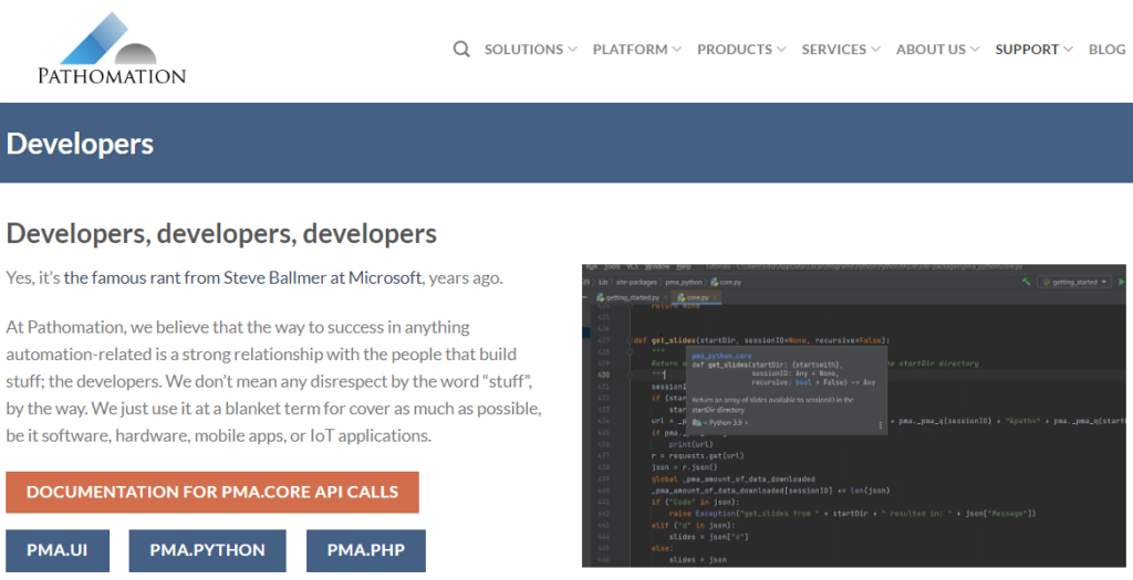
Middleware
At the end of the day, what we do is straightforward: Pathomation makes middleware software for digital pathology.
Now depending on who you talk to, one or more terms in that statement may take some explaining:
- Pathomation: it’s not phantomation (ghostbusters, anybody?), photomation (photonics, quantum physics; nah, to be honest we’re probably not smart enough)… It’s Pathomation, from Pathology – Automation.
- Middleware software: Middleware acts as a broker between different components. Your typical example of middleware would be a printer driver, which allows a user to convert text and pixels from the computer screen to ink on paper. On that note, “pixel broker”, is another way how we like to describe ourselves. The slide scanner converts the tissue from the glass slide into a (huge!) collection of pixels, and we make sure these pixels can be read and presented properly, regardless of what scanner was used to generate them, and regardless of where they are stored
- Digital pathology: it started in the 60s at Massachusetts General Hospital in Boston with tele-pathology. In the 2000s, engineers figured out that they could automate microscopes to take sequential pictures of various regions of interest on glass slides, and then stitch those to create (again: huge!) compound images that would let pathologists bypass the traditional microscope altogether and use the computer screen as a virtual microscope.
- Pathology: the medical sub-specialty that diagnoses disease at the smallest level. Up to 70% of therapeutic treatment is attributable to one pathological exam or another. That’s… huge actually, and it’s why it’s all the more important to make the pixels from the scan flow to that storage location to that computer screen as smooth as possible.
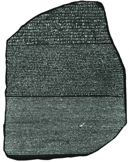
But here’s another way of looking at is: Pathomation is the Rosetta Stone to your digital pathology content.
We make middleware software that optimizes the transport of pixels. We show the pathologist the pixels he or she needs, when he or she wants, where he or she desires to have them.
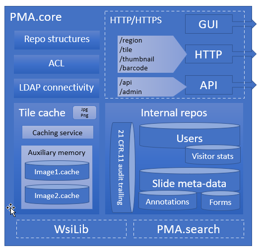
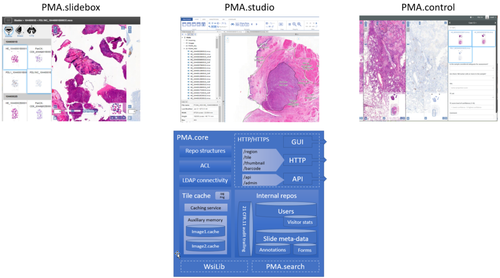
The central piece of software we develop for that is called PMA.core, and on top of PMA.core we have a variety of end-user front-end applications like PMA.slidebox, PMA.studio, or PMA.control.
Growing software
We didn’t start off with these though. So bear with us as we take a little trip down memory lane.
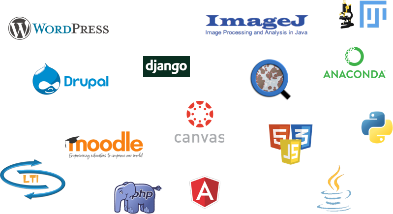
Once you have a successful piece of software, it doesn’t take long for people to ask “hey, can I also use it for this (or that)?”. Therefore, on top of PMA.core, we built several Software Development Kits (SDKs) that make it easier for other software developers to integrate digital pathology into their own applications (image analysis (AI/DL/ML), APLIS, LIMS, PACS…).
The next question is: “I don’t know how to program. I just have a WordPress blog that I want to embed some slides in.” So we wrote a series of plugins for various Content Management Systems (CMS), Learning Management Systems (LMS), and even third-party Image Analysis (IA) tools.
Eventually, we got into integrated end-user application development, too. As far as we’re concerned, we have three software packages that cater to end-users:
- PMA.slidebox caters to educational applications whereby people just want to share collections with students, conference participants, or peers. A journal could benefit from this and publish virtual slides via a slidebox to serve as supplemental material to go with select author papers.
- PMA.studio wants to be a pathologist’s cockpit. You can have slides presented in grid layouts, coming from different PMA.core servers. But if you’re an image analyst working with whole slide images (WSI), that works, too. Integrate data sources, and annotations. Have a video conference, and prepare high-resolution images (snapshots) for your publications… Do it all from the convenience of a single flexible and customizable user interface. If you’re an oncologist or surgeon that only occasionally wants to look along, PMA.studio may be a bit of overkill. But to build custom portals for peripheral users, you have those SDKs of course.
- PMA.control goes above and beyond the simple collections that you place online with PMA.slidebox. With PMA.control, you can manage participants, manage what content they see at that what time, organize complex training sessions, organize the development and evaluation of new scoring schemes etc. With PMA.control, you are in… control.

A platform
Pathomation solves one problem really well. Because the problem manifests itself in many ways, we offer a number of routes to address it.
Different scanners output different file formats, that need to be distributed in different ways.
Due to the diverse applications of virtual microscopy and digital pathology, we conceived our own toolbox different from the beginning. Instead of a single application, we set off from the start to build a platform instead.
Pathomation offers a software platform.
- You start with PMA.core, which handles virtual slides, user management, data capture, and audit trailing. All applications need these capabilities.
- On top of PMA.core comes a range of connectable components. These can take the form of an application (desktop, web, mobile), but can also be connecting pieces to other external software. We do not offer Image Analysis, but we give you the right connectors to pass your data on to your favorite AI environment. We do not have our own LIMS, but we offer technical building blocks (APIs) for independent vendors to embed slide handling capabilities on their end on top of our central PMA.core.
So whether you’re looking to build custom portals for your own user-base (PMA.UI framework), or are looking for a one-stop solution to evaluate new histological scoring protocols (PMA.control); we have you covered.